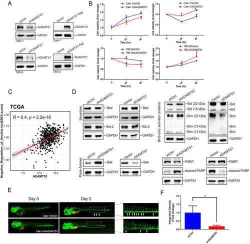
ADAMTS1 expression promotes anoikis resistance of renal cell carcinoma (RCC) via inhibiting Bid, Bim, and Bak. A A western blot analysis was conducted to assess ADAMTS1 expression in Caki-1 and 786-O cells following transduction with either ADAMTS1 short hairpin (sh)RNA (left) or an ADAMTS1-expressing vector (right). B Cell viability of suspended Caki-1 and 786-O cells was evaluated using a CCK8 assay at 24 and 48 h post-stable overexpression (right) or knockdown (left) of ADAMTS1. Data are presented as the mean ± SD of three independent experiments. * p < 0.05, compared with the control group. C Dot plot demonstrated the correlation between the single sample gene set enrichment analysis (ssGSEA) score of “negative regulation of anoikis” and ADAMTS1 expression in TCGA-KIRC patients. A Pearson correlation was performed to evaluate their association and significance. D Western blot analysis of intrinsic apoptosis-related proteins (Bad, Bcl-2, Bak, Bid, Bim, and PARP) in suspended Caki-1 cells manipulated with ADAMTS1. GAPDH served as a loading control. E and F Dissemination of RCC cells in zebrafish embryos. Caki-1 cells with ADAMTS1-knockdown were implanted into zebrafish embryos at 48 h post fertilization. Tumor cell dissemination was observed at 2 days post injection (dpi), with disseminated tumor foci indicated by white arrowheads on the trunk and end-tail (E). Integrated densities of Caki-1 metastatic tumor cells in the zebrafish trunk and end-tail at 2 dpi were quantified, with the mean value of the integrated density in the shCtl group set to onefold (F)
|
