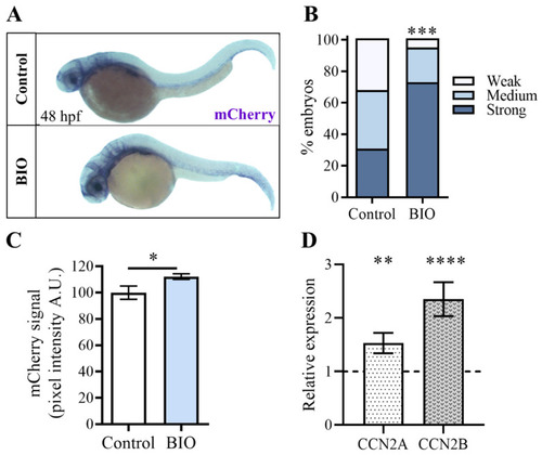
Pharmacological inhibition of β-catenin kinase GSK3 increases Tg(Hsa.CTGF:nlsmCherry)ia49 reporter signal. (A) Tg(Hsa.CTGF:nlsmCherry)ia49 embryos were exposed to BIO from 24 to 48 hpf, and after fixation, the mCherry transgene expression was assessed by in situ hybridization. (B,C) The embryos treated with BIO showed higher levels of mRNA mCherry transgene expression compared to their untreated control siblings. In the in situ hybridization experiments, the signal intensity variation was assessed using two different methods. In (B), the analysis was performed by grouping the embryos into three distinct classes based on the observed signal strength at the whole embryo level. In (C), the mCherry signals were quantified by measuring the pixel intensity in the head region, using Volocity 6.0 software. Control (n = 52), BIO (n = 51). (D) In 48 hpf zebrafish embryos, the expression of ccn2a and ccn2b genes was strongly up-regulated after treatment with BIO for 24 h. mRNA levels were normalized to the DMSO-treated controls (dashed line). Scale bar: 250 μm. In (C,D), data are presented as mean +/− SEM (Mann−Whitney test in (C) and Student t-test in (D); * p < 0.05, ** p < 0.01, *** p < 0.001, **** p < 0.0001).
|
