|
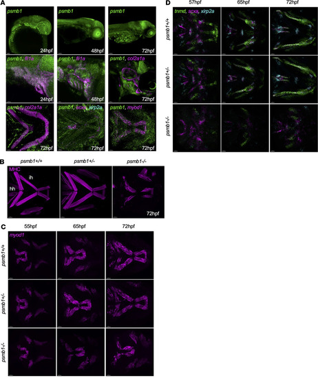
Craniofacial muscle and tendon defects in psmb1 mutants. (A) RNAscope to assess developmental timing and distribution of psmb1 expression. Top: psmb1 (green) expression at 24, 48, and 72 hpf. Scale bars: 50 μm, 100 μm, and 100 μm, respectively. Middle: psmb1 expression in developing pharyngeal arches and cartilage. Left and middle: psmb1 (green), fli1a (magenta), right: psmb1 (green), col2a1a (magenta). Scale bars: 50 μm. Bottom: psmb1 expression in craniofacial tissues at 72 hpf. Left: psmb1 (green), col2a1a (magenta). Middle: psmb1 (green), myod1 (magenta). Right: psmb1 (green), scxa (magenta), xirp2a (cyan). Scale bars: 30 μm. (B) Antibody stain for myosin heavy chain (MHC) at 72 hpf showing defects in craniofacial muscles in psmb1 mutants. Scale bars: 50 μm. hh, hyohyal; ih,interhyal. (C) RNAscope for myod1 at 55–72 hpf. Scale bars: 30 μm. (D) RNAscope for tnmd (green), scxa (magenta), and xirp2a (cyan) at 57–72 hpf demonstrating defects in tendon development in psmb1 mutants. Scale bars: 30 μm.
|