IMAGE

Figure 5

ID
ZDB-IMAGE-240826-7
Genes
Source
Figures for Miller et al., 2024
Image
Figure Caption

Figure 5 
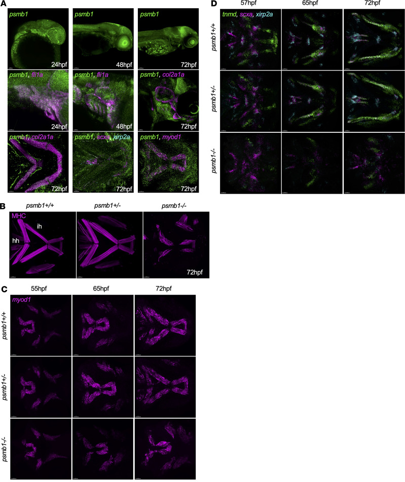
Craniofacial muscle and tendon defects in psmb1 mutants.

(A) RNAscope to assess developmental timing and distribution of psmb1 expression. Top: psmb1 (green) expression at 24, 48, and 72 hpf. Scale bars: 50 μm, 100 μm, and 100 μm, respectively. Middle: psmb1 expression in developing pharyngeal arches and cartilage. Left and middle: psmb1 (green), fli1a (magenta), right: psmb1 (green), col2a1a (magenta). Scale bars: 50 μm. Bottom: psmb1 expression in craniofacial tissues at 72 hpf. Left: psmb1 (green), col2a1a (magenta). Middle: psmb1 (green), myod1 (magenta). Right: psmb1 (green), scxa (magenta), xirp2a (cyan). Scale bars: 30 μm. (B) Antibody stain for myosin heavy chain (MHC) at 72 hpf showing defects in craniofacial muscles in psmb1 mutants. Scale bars: 50 μm. hh, hyohyal; ih,interhyal. (C) RNAscope for myod1 at 55–72 hpf. Scale bars: 30 μm. (D) RNAscope for tnmd (green), scxa (magenta), and xirp2a (cyan) at 57–72 hpf demonstrating defects in tendon development in psmb1 mutants. Scale bars: 30 μm.

Figure Data
Acknowledgments
This image is the copyrighted work of the attributed author or publisher, and ZFIN has permission only to display this image to its users. Additional permissions should be obtained from the applicable author or publisher of the image. Full text @ JCI Insight