|
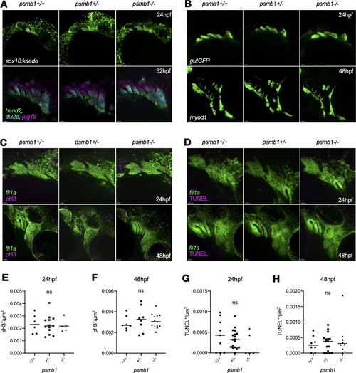
psmb1 is dispensable for pharyngeal arch morphogenesis and patterning. (A) Imaging of sox10:kaede embryos at 24 hpf (top, scale bars: 50 μm) did not reveal any defects with neural crest cell migration to the arches. RNAscope for hand2, dlx2a, and jag1b demonstrates normal dorsal-ventral arch patterning in psmb1 mutants at 32 hpf (bottom, scale bars: 30 μm). (B) Imaging of gutGFP embryos at 24 hpf and myod1 RNAscope at 48 hpf demonstrates that the pharyngeal arch endoderm and muscle are formed normally in psmb1 mutants. Scale bars: 50 μm. (C) pH3 antibody staining (magenta) in fli1a:EGFP embryos at 24 hpf and 48 hpf to assess neural crest cell proliferation in psmb1 mutants. Scale bars: 30 μm (D) TUNEL staining (magenta) in fli1a:EGFP embryos at 24 hpf and 48 hpf to assess neural crest cell death in psmb1 mutants. Scale bars: 20 μm. (E and F) Quantification of proliferation in pharyngeal arches 1 and 2 in images in C; normalization is to arch 1 and 2 fli1a+ area. (E): n = 6 (+/+), 13 (+/–), 5 (–/–). (F): n = 7 (+/+), 8 (+/–), 13 (–/–). (G and H) Quantification of cell death in pharyngeal arches 1 and 2 in images in D; normalization is to fli1a+ arch 1 and 2 area. (G): n = 9 (+/+), 18 (+/–), 5 (–/–). (H): n = 8 (+/+), 17 (+/–), 8 (–/–). Data shown represent mean ± SD. Significance was calculated with 1-way ANOVA with Dunnett’s multiple comparisons test, ns: not significant.
|