
Fig. 7
- ID
- ZDB-FIG-240807-7
- Publication
- Fuentes et al., 2024 - Maternal regulation of the vertebrate oocyte-to-embryo transition
- Other Figures
- All Figure Page
- Back to All Figure Page
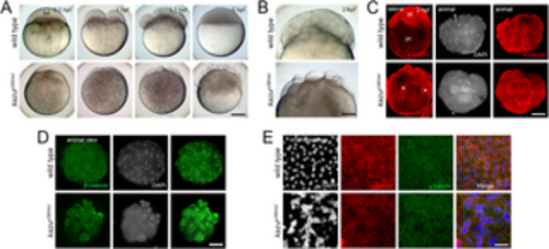
Phenotypic characterization of the kazup26thbd mutant early embryo. A. Bright field images showing early developmental stages of whole-mount live wild-type (n = 99/99 from 2 females) and kazup26thbd (n = 185/265 from 2 females) embryos. The mutant phenotype reveals a smaller blastodisc (bd) with smaller cells during cleavage. These abnormalities in cell size acquisition, together with asynchronous and unequal cell divisions, leads to the formation of a defective blastula with many detached blastomeres. B. Higher magnification images showing a cellularized wild-type blastoderm (n = 125/125). In contrast, kazup26thbd embryos exhibit a less compacted blastoderm containing smaller cells (n = 62/91). C. Lateral and animal views of whole-mount wild-type (n = 31/35) and mutant (n = 39/47) early embryo that show the distribution of α-Tubulin at 2 hpf. In wild-type embryos, α-Tubulin is mainly distributed in the blastoderm (bt) and dividing blastomeres. In kazup26thbd mutants, α-Tubulin is distributed in different sized cells and throughout the yolk cell (yc, asterisks). D. Animal views of whole-mount wild-type (n = 26) and mutant (n = 8) early embryos that show the localization of β-Catenin at 2 hpf. In wild-type embryos, β-Catenin localized to the blastomere boundaries. In kazup26thbd mutants, β-Catenin is aberrantly localized in different cytoplasmic domains. E. Animal views of DAPI, α-tubulin, and centrosome staining showing abnormal blastoderm formation at the sphere stage. Cell boundaries and mitotic figures are not observed in most of the mutant (n = 25/33) compared to wild-type (n = 30/35) embryos examined. Notice the formation of a syncytial nuclei layer in the 5 hpf kazup26thbd embryo. Scale bar = 180 μm (A), 110 μm (B), 230 μm (C), 165 μm (D), 100 μm (E). |