
Fig. 4
- ID
- ZDB-FIG-240807-4
- Publication
- Fuentes et al., 2024 - Maternal regulation of the vertebrate oocyte-to-embryo transition
- Other Figures
- All Figure Page
- Back to All Figure Page
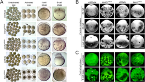
Egg activation mutants. A. Bright-field images showing early developmental stages of live wild-type and mutant eggs and embryos. All unactivated eggs collected after gently squeezing gravid females showed no detectable defects. After egg activation, however, the krang mutant egg displays a pronounced chorion elevation defect (black arrows). As development proceeds, the cytoplasm is abnormally distributed in the yolk cell (yc) of spotty mutant eggs and early embryos (black arrowheads) and a defective blastoderm (bt) is formed. krang (1887 eggs from 50 females), spotty (1273 eggs from 40 females), and p28tabj (2506 eggs from 34 females) and kazu (1608 eggs from 32 females). B. Lateral view of acid-fixed wild-type and mutant eggs and early embryos showing the organization of the central cytoplasm (bright). In wild type, the blastodisc (bd) grows by animal-ward transport of cytoplasm from the yolk cell (yc) and by the 4-cell stage, the cytoplasm in the yolk cell is no longer evident. In contrast, spotty, p28tabj and kazu mutant eggs exhibited an abnormal distribution and severe retention of cytoplasm in the yolk cell (black asterisks). Wild type (n = 22), spotty (n = 17), and p28tabj (n = 19) and kazu (n = 12). C. Lateral view of acid-fixed and DiOC6-stained wild-type and mutant eggs and early embryos showing the organization of the cortical cytoplasm (green) and yolk (dark). Wild type (n = 20), spotty (n = 20), and p28tabj (n = 14) and kazu (n = 18). Scale bar = 830 μm (A, columns 1 and 2 from left to right), 150 μm (A, columns 3 and 4 from left to right), 190 μm (B, C). |