
Fig. 1 Supplemental 3
- ID
- ZDB-FIG-240729-34
- Publication
- Weiss et al., 2024 - A syngeneic spontaneous zebrafish model of tp53-deficient, EGFRvIII, and PI3KCAH1047R-driven glioblastoma reveals inhibitory roles for inflammation during tumor initiation and relapse in vivo
- Other Figures
- All Figure Page
- Back to All Figure Page
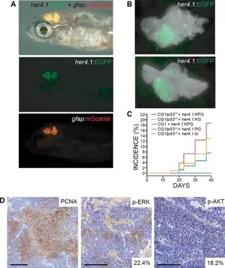
her4.1-driven over-expression of KRASG12D + PI3KCAH1047R drives glial-derived brain tumor formation in syngeneic tp53 loss-of-function mutant zebrafish. (A) her4.1:mScarlet and gfap:GFP expression in the anterior CNS of 30 days post fertilization (dpf) syngeneic (CG1) tp53-/- zebrafish injected at the one-cell stage with linearized her4.1:KRASG12D + her4.1:PI3KCAH1047R + her4.1:EGFP + gfap:mScarlet transgenes. (B) Whole brain dissected from mosaic-injected her4.1:KRASG12D + her4.1:PI3KCAH1047R + her4.1:EGFP zebrafish at 30 dpf. (C) Cumulative frequencies of EGFP+ brain lesions in syngeneic tp53-/- mutant (CG1tp53-/-) and wild-type (CG1) zebrafish injected at the one-cell stage with her4.1:KRASG12D (K), her4.1:PI3KCAH1047R (P), and/or her4.1:EGFP (G). (D) Immunohistochemical staining of proliferating cell nuclear antigen (PCNA), phosphorylated-ERK (p-ERK), and phosphorylated-AKT (p-AKT) on her4.1:KRASG12D + her4.1:PI3KCAH1047R + her4.1:EGFP tumor section. Quantification of p-ERK and p-AKT positive cells within total field. Scale bars represent 50 μm. |