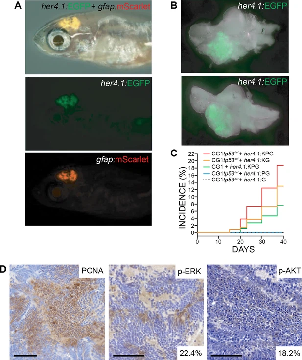
Fig. 1 Supplemental 3 her4.1-driven over-expression of KRASG12D + PI3KCAH1047R drives glial-derived brain tumor formation in syngeneic tp53 loss-of-function mutant zebrafish. (A) her4.1:mScarlet and gfap:GFP expression in the anterior CNS of 30 days post fertilization (dpf) syngeneic (CG1) tp53-/- zebrafish injected at the one-cell stage with linearized her4.1:KRASG12D + her4.1:PI3KCAH1047R + her4.1:EGFP + gfap:mScarlet transgenes. (B) Whole brain dissected from mosaic-injected her4.1:KRASG12D + her4.1:PI3KCAH1047R + her4.1:EGFP zebrafish at 30 dpf. (C) Cumulative frequencies of EGFP+ brain lesions in syngeneic tp53-/- mutant (CG1tp53-/-) and wild-type (CG1) zebrafish injected at the one-cell stage with her4.1:KRASG12D (K), her4.1:PI3KCAH1047R (P), and/or her4.1:EGFP (G). (D) Immunohistochemical staining of proliferating cell nuclear antigen (PCNA), phosphorylated-ERK (p-ERK), and phosphorylated-AKT (p-AKT) on her4.1:KRASG12D + her4.1:PI3KCAH1047R + her4.1:EGFP tumor section. Quantification of p-ERK and p-AKT positive cells within total field. Scale bars represent 50 μm.
Image
Figure Caption
Acknowledgments
This image is the copyrighted work of the attributed author or publisher, and
ZFIN has permission only to display this image to its users.
Additional permissions should be obtained from the applicable author or publisher of the image.
Full text @ Elife

