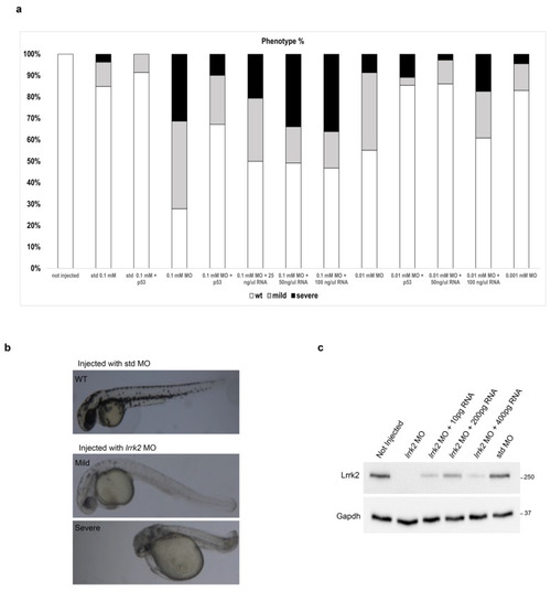
Effects of lrrk2 MO on zebrafish embryo development. (a) Analysis of morphological phenotypes of zebrafish embryos injected with lrrk2 MO (0.1 mM–0.001 mM), 0.1 mM std MO, lrrk2 MO (0.1 mM–0.001 mM) + 0.1 mM p53 MO, lrrk2 MO (0.1 and 0.01 mM) + human LRRK2 mRNA (25, 50, and 100 ng/μL corresponding to 100, 200, and 400 pg). Quantification of the absolute percentage of embryos showing the wild-type-like (white closed bars), mild (gray closed bars), and severe (black closed bars) morphological phenotypes out of the total embryos analyzed. (b) Representative images of the phenotypes observed after the injection with std MO and lrrk2 MO. Number of observed embryos: not injected (n = 350); 0.1 mM std MO (n = 65); 0.1 mM std MO + p53 MO (n = 60); 0.1 mM lrrk2 MO (n = 230); 0.01 mM lrrk2 MO (n = 196); 0.001 mM lrrk2 MO (n = 112); 0.1 mM lrrk2 MO + 0.1 mM p53 MO (n = 131); 0.01 mM lrrk2 MO + 0.1 mM p53 MO (n = 110); 0.1 mM lrrk2 MO + 25 ng/μL hLRRK2 mRNA (n = 44); 0.1 mM lrrk2 MO + 50 ng/μL hLRRK2 mRNA (n = 57); 0.1 mM lrrk2 MO + 100 ng/μL hLRRK2 mRNA (n = 47); 00.1 mM lrrk2 MO + 50 ng/μL hLRRK2 mRNA (n = 56); 0.01 mM lrrk2 MO + 100 ng/μL hLRRK2 mRNA (n = 53). (c) A pool of fifty embryos per condition (not injected, lrrk2 MO, lrrk2 MO + 100 pg hLRRK2 mRNA, lrrk2 MO + 200 pg hLRRK2 mRNA, lrrk2 MO + 400 pg hLRRK2 mRNA, std MO) were subjected to immunoblotting using Lrrk2 and Gapdh antibodies at 48 hpf.
|
