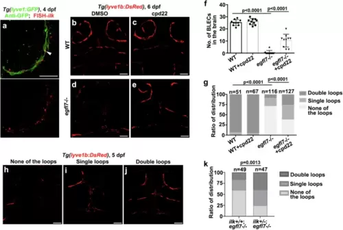
Egfl7-Integrin αvβ3 plays a role in BLECs formation through ILK pathway.a FISH and antibody staining shows ilk is expressed in GFP+ BLECs at 4 dpf (arrowheads). n = 17/18 embryos. b–g Inhibition of ILK by cpd22 can partially rescue the absence of BLECs in the egfl7 mutant at 6 dpf (b–e). Quantification of the number of BLECs in the double loops of brain in different treatment groups (f, n = 10 embryos, two-tailed unpaired t test. Data are represented as mean ± SD). The statistics show the percentage of embryos that have double lymphatic loops, single loops, and none of the loops in the brain (g, WT, n = 51 embryos, WT + cpd22, n = 67 embryos, egfl7-/-, n = 116 embryos, egfl7-/- + cpd22, n = 127 embryos, χ2 test).). h–k An egfl7 mutant was crossed with an ilk heterozygote to generate two types of larvae: ilk+/+; egfl7-/- and ilk+/-; egfl7-/-, which were then studied for BLECs loop-structures formation. The larvae were classified into three categories based on their phenotype: Double loops, single loops, and none of the loops (h–j). The results showed that compared to ilk+/+; egfl7-/-, the ilk+/-; egfl7-/- larvae had a decreased percentage of the none of the loops phenotype, and a partially rescued percentage of double loops and single loops phenotype (k, ilk+/+; egfl7-/-, n = 49 embryos, ilk+/-; egfl7-/-, n = 47 embryos, χ2 test). Scale bar, 50 μm.
|
