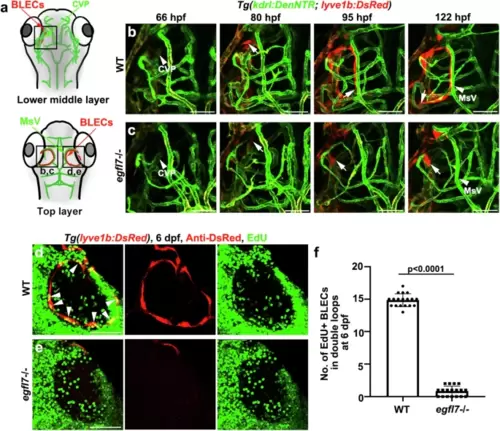
egfl7 is essential for the migration and proliferation of BLECs.a Schematic diagram showing the lower middle layer and top layer of the vessels in the brain, respectively. Black frames indicate the image area of corresponding panels. CVP (choroidal vascular plexus) is observed in the lower middle layer; MsV (mesencephalic vein) is in the top layer. b Time-lapse image of Tg(lyve1b: DsRed; kdrl: DenNTR) from 66 hpf to 122 hpf show the BLECs (arrows) sprout from CVP (arrowhead at 66 hpf) and migrate along the MsV (arrowhead) to form the lymphatic loop in WT at 122 hpf. n = 15 embryos. c The BLECs (arrows) of egfl7 mutant shows a delayed sprouting from CVP (arrowhead at 66 hpf), fail to migrate along the MsV (arrowhead), and is unable to form the loop at 122 hpf. n = 15 embryos. d–f EdU staining shows the proliferation of BLECs in WT and egfl7 mutant at 6 dpf, the EdU is injected at 56 hpf. Note the EdU+ BLECs is significantly decreased in the mutant (d, e). Arrowheads indicate the EdU+ BLECs in WT. The statistics show the number of EdU+ BLECs in double loops at 6 dpf in WT and egfl7 mutant (f, n = 18 embryos; two-tailed unpaired t-test). Data are represented as mean ± SD. Scale bar, 50 μm.
|
