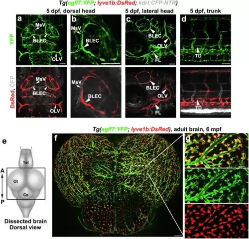
Tg(egfl7:YFP) transgenic zebrafish express YFP in blood vessels and lymphatics.a–d Tg(egfl7:YFP) was generated showing YFP in blood vessels and lymphatics. The distribution of YFP is similar with CFP and DsRed in the triple transgenic line Tg(egfl7:YFP; lyve1b: DsRed; kdrl: CFP-NTR) at 5 dpf. n = 25/25 embryos. The low-magnified image shows the dorsal view of the head (a), the high-magnified image in another embryo shows the magnified BLECs loop in the dorsal brain (b). The lateral facial lymphatics of the head shows in c. The trunk lymphatics shows in d. BLECs, brain lymphatic endothelial cells, MsV, mesencephalic vein, OLV, otolithic lymphatic vessel, FL, facial lymphatics, TD, thoracic duct. Scale bar, 50 μm. e Schematic diagram showing the dorsal view of a dissected adult zebrafish brain. f egfl7 promoter-driven YFP is expressed in all BLECs and blood vessels. n = 6/8 adults. g High-magnification inset showing the YFP overlap with DsRed in the double transgenic line Tg(egfl7:YFP; lyve1b: DsRed). Scale bars, 200 μm in f and 50 μm in g.
|
