
Fig. 2
- ID
- ZDB-FIG-240702-227
- Publication
- Wang et al., 2020 - Key Developmental Regulators Suggest Multiple Origins of Pancreatic Beta Cell Regeneration
- Other Figures
- All Figure Page
- Back to All Figure Page
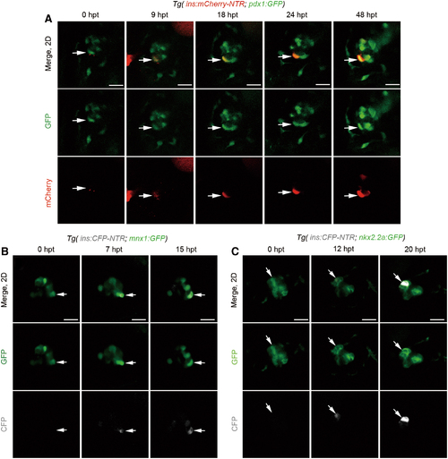
Nascent β cells emerged from pre-existing pdx1+, mnx1+, or nkx2.2a+ cells. (A) A set of real-time imaging of a regenerating β cell (white arrows) in the primary islet after Mtz treatment using the Tg(ins:mCherry-NTR; pdx1:GFP) transgenic line (n = 19/23). Merged and single-channel confocal planes showed a pdx1+ins- cell (0 hpt, white arrow) committed to the β cell fate in the next 48 h. β cell maturation was noted by the onset of mCherry fluorescence indicating insulin expression since 9 hpt. mCherry expression was getting steadily stronger from 18 to 48 hpt, indicating the maintenance of β cell identity. (B, C) Time course of a regenerating β cell (white arrows) in the primary islet after Mtz treatment using the Tg(mnx1:GFP; ins:CFP-NTR) or Tg(nkx2.2a:GFP; ins:CFP-NTR), respectively. A GFP+CFP− cell (white arrow) showed commitment to the β cell fate within several hours after removal of Mtz (B, n = 21/25; C, n = 18/26; scale bar, 20 μm). Color images are available online. |