
Fig. 1
- ID
- ZDB-FIG-240702-226
- Publication
- Wang et al., 2020 - Key Developmental Regulators Suggest Multiple Origins of Pancreatic Beta Cell Regeneration
- Other Figures
- All Figure Page
- Back to All Figure Page
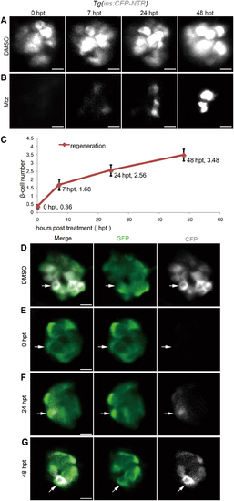
β cell regeneration after a near-total ablation. (A, B) Representative 3D projections of β cells in the primary islet of Tg(ins:CFP-NTR) larvae from 0 to 48 hpt. The larvae treated with DMSO in the control group showed steady expression of CFP in β cells of the primary islet (A, n = 30/30; scale bar, 10 μm). The larvae treated with Mtz showed 0 β cell at 0 hpt. Nascent β cells emerged as regeneration proceeded (B, n = 32/37; scale bar, 10 μm). (C) Quantification of nascent β cells at different time points post-treatment. The mean values of β cell number are indicated (n = 25 for each time point. Data are expressed as mean ± s.e.m.). (D–G) Time series of merged and single-channel confocal planes of primary islets treated with DMSO or Mtz. Tg(ins:CFP-NTR) was used to label β cells blue with CFP and perform ablation. Tg(neurod:EGFP) marked neurod+ cells with GFP. All β cells were neurod-positive in both the DMSO-treated control group and the Mtz-treated group. A neurod+ins- cell at 0 hpt showed positive CFP signal in the following 48 h (white arrows) indicating its commitment to β cell fate (D, n = 23/23; E, n = 17/19; scale bar, 10 μm). 3D, three-dimensional; CFP, cyan fluorescent protein; DMSO, dimethyl sulfoxide; GFP, green fluorescent protein; Mtz, metronidazole; NTR, nitroreductase; s.e.m., standard error of the mean. Color images are available online. |