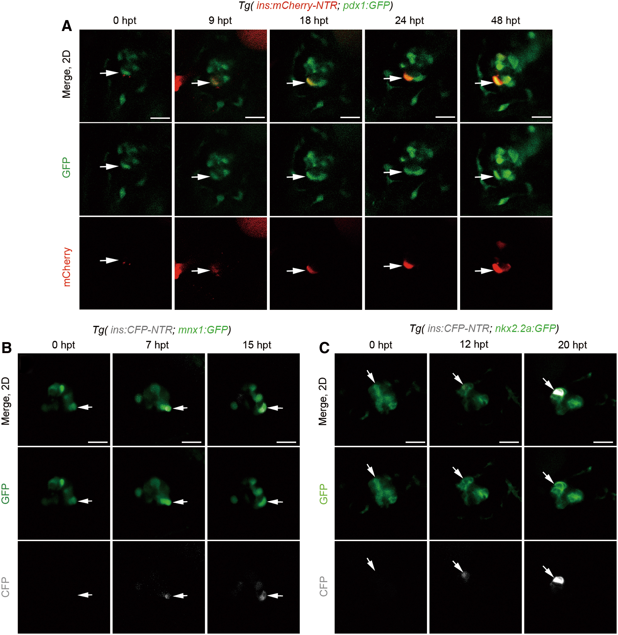
Fig. 2 Nascent β cells emerged from pre-existing pdx1+, mnx1+, or nkx2.2a+ cells. (A) A set of real-time imaging of a regenerating β cell (white arrows) in the primary islet after Mtz treatment using the Tg(ins:mCherry-NTR; pdx1:GFP) transgenic line (n = 19/23). Merged and single-channel confocal planes showed a pdx1+ins- cell (0 hpt, white arrow) committed to the β cell fate in the next 48 h. β cell maturation was noted by the onset of mCherry fluorescence indicating insulin expression since 9 hpt. mCherry expression was getting steadily stronger from 18 to 48 hpt, indicating the maintenance of β cell identity. (B, C) Time course of a regenerating β cell (white arrows) in the primary islet after Mtz treatment using the Tg(mnx1:GFP; ins:CFP-NTR) or Tg(nkx2.2a:GFP; ins:CFP-NTR), respectively. A GFP+CFP− cell (white arrow) showed commitment to the β cell fate within several hours after removal of Mtz (B, n = 21/25; C, n = 18/26; scale bar, 20 μm). Color images are available online.
Image
Figure Caption
Acknowledgments
This image is the copyrighted work of the attributed author or publisher, and
ZFIN has permission only to display this image to its users.
Additional permissions should be obtained from the applicable author or publisher of the image.
Full text @ Zebrafish

