
Fig. 4
- ID
- ZDB-FIG-240702-229
- Publication
- Wang et al., 2020 - Key Developmental Regulators Suggest Multiple Origins of Pancreatic Beta Cell Regeneration
- Other Figures
- All Figure Page
- Back to All Figure Page
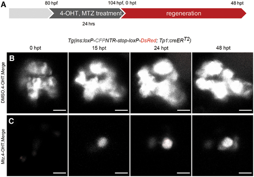
Pancreatic ductal cells did not contribute to nascent β cells. (A) The strategy for Cre/loxP-mediated lineage tracing of pancreatic ductal cells in β cell regeneration. 4-OHT was used in both the DMSO-treated control group and Mtz-treated group to label Notch-responsive pancreatic ductal cells. Mtz was used only in the Mtz-treated group, but not in the DMSO-treated control group. (B, C) 3D projections of islets in control and Mtz treatment groups from 0 to 48 hpt using Tg(Tp1:creERT2; ins:loxP-CFPNTR-stop-loxP-DsRed) transgenic lines. Notch-responsive pancreatic ductal cells should be labeled by DsRed after 4-OHT treatment. Data showed no β cell was positive for DsRed in control group (B, n = 19/19). Nascent β cells were not positive for DsRed, indicating a negative contribution of tp1+ pancreatic ductal cells to the nascent β cells (C, n = 18/23). Three tp1+ red fluorescent cells showed in the (C) 0 hpt were dying/dead β cells with tp1 activated during Mtz treatment. They disappeared thereafter possibly due to cell death. Scale bar, 10 μm. 4-OHT, 4-hydroxy-tamoxifen. Color images are available online. |