FIGURE

Figure 3

ID
ZDB-FIG-240613-44
Publication
D'Gama et al., 2024 - Ciliogenesis defects after neurulation impact brain development and neuronal activity in larval zebrafish
Other Figures
All Figure Page
Back to All Figure Page
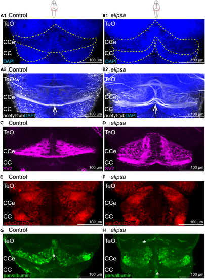
Figure 3

Cerebellar defects in 4 days old elipsa mutants

(A1–B1) Cell nucleus stained using dapi in control (A1) and elipsa mutant (B1). Differences in morphology indicated by yellow lines. n = 7 controls and 7 mutants. (A2–B2) Larvae immunostained with anti-acetylated tubulin to stain axonal bundles (indicated with white arrow) in control (A2) and elipsa mutant (B2). n = 7 controls and 7 mutants.

(C and D) Larvae immunostained with the presynaptic vesicle marker SV2 in control (C) and elipsa mutant (D). n = 6 controls and 6 mutants (E and F) Expression of glutamatergic cells in Tg(vglut2a:dsRed) transgenic zebrafish in control (E) and elipsa mutant (F). n = 8 controls and 8 mutants (G and H) Immunostaining of cerebellar Purkinje neurons using anti-parvalbumin antibody in control (G) and elipsa mutant (H). n = 10 controls and 8 mutants. ∗ indicates nonspecific signal from blood cells and vasculature. Number of Purkinje cells in control (mean ± standard deviation): 83.87 ± 5.22 cells, in mutants: 58 ± 6.8 cells, p-value 4,6965E-06 according to Student’s t test. Teo, Optic Tectum; CCe, Corpus Cerebelli; CC, Crista cerebralis.

Expression Data

Expression Detail
Antibody Labeling
Phenotype Data

Phenotype Detail
Acknowledgments
This image is the copyrighted work of the attributed author or publisher, and ZFIN has permission only to display this image to its users. Additional permissions should be obtained from the applicable author or publisher of the image. Full text @ iScience