
Figure 3
- ID
- ZDB-IMAGE-240613-44
- Genes
- Antibodies
- Publication
- D'Gama et al., 2024 - Ciliogenesis defects after neurulation impact brain development and neuronal activity in larval zebrafish
- All Figures
- Figures for D'Gama et al., 2024
|
Figure 3
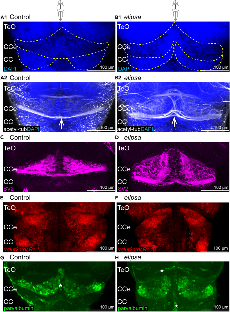
Cerebellar defects in 4 days old
(A1–B1) Cell nucleus stained using dapi in control (A1) and
(C and D) Larvae immunostained with the presynaptic vesicle marker SV2 in control (C) and