FIGURE

Figure 1

ID
ZDB-FIG-240613-42
Publication
D'Gama et al., 2024 - Ciliogenesis defects after neurulation impact brain development and neuronal activity in larval zebrafish
Other Figures
All Figure Page
Back to All Figure Page
Figure 1

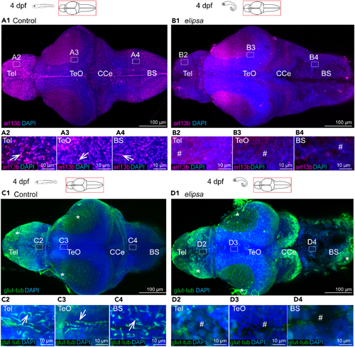
Loss of primary and motile cilia in the elipsa mutant larval brain

(A1–A4 and B1–B4) Staining of dissected 4 dpf brains with arl13b antibody to stain all cilia in the brain, n = 8 controls and 9 mutants. A1 At 4 dpf, arl13b stained cilia were located all over the brain, represented in insets drawn in the telencephalon (A2), optic tectum (A3) and brainstem (A4).

(B1) In the elipsa mutants, arl13b-expressing cilia were absent in the entire brain, as represented by insets drawn in the telencephalon (B2), optic tectum (B3), and brain stem (B4). (C1–C4 and D1–D4) Staining of dissected 4 dpf brains with glutamylated tubulin used to identify motile cilia, n = 5.

(C1) At 4 dpf, single glutamylated tubulin-positive cilia were present in the forebrain choroid plexus (C2), on the dorsal roof and ventral part (C3) of the tectal/diencephalic ventricle and in the rhombencephalon choroid plexus (C4).

(D1) In elipsa mutant, glutamylated tubulin-positive cilia were absent in the telencephalon (D2), optic tectum (D3), and brain stem (D4). Tel, Telencephalon; Teo, Optic Tectum; CCe, Corpus Cerebelli; BS, Brain stem. Cilia loss indicated by # symbol and nonspecific signal from glutamylated tubulin is represented by ∗ symbol.

See also Figures S1 and S2.

Expression Data

Expression Detail
Antibody Labeling
Phenotype Data

Phenotype Detail
Acknowledgments
This image is the copyrighted work of the attributed author or publisher, and ZFIN has permission only to display this image to its users. Additional permissions should be obtained from the applicable author or publisher of the image. Full text @ iScience