|
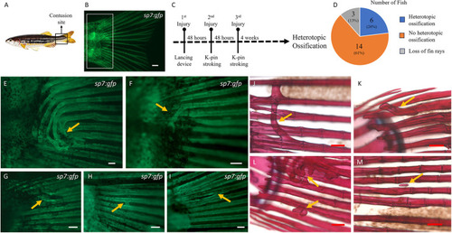
Heterotopic bone formation at the tail fin lepidotrichia. (A) Site of caudal peduncle contusion (boxed area). (B) Appearance of an uninjured tail fin under fluorescence microscopy in a transgenic Tg(sp7:gfp) zebrafish. The boxed area represents the site where heterotopic bone formation was noted, as shown in E-M. (C) Timeline representing the recurring injuries with a 48-h interval in between, followed by a 4-week wait to observe heterotopic bone. (D) Pie chart showing the overall number of fish with and without heterotopic bone (n=23). (E-I) Fluorescence microscopy of injured Tg(sp7:gfp) zebrafish. (J-M) Alizarin Red staining of injured wild-type zebrafish. Exuberant heterotopic bone formation (arrow in E) bridging multiple adjacent fin rays beneath the overlying soft tissues of the caudal peduncle and various other forms of heterotopic bone (arrows in F-M) were observed. Scale bars: 500 µm (B,E); 200 µm (F-M).
|