
Fig. 7
- ID
- ZDB-FIG-240523-69
- Publication
- Akbari et al., 2024 - Label-free, whole-brain in vivo mapping in an adult vertebrate with third harmonic generation microscopy
- Other Figures
- All Figure Page
- Back to All Figure Page
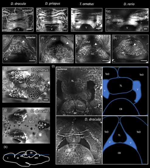
Comparisons of third harmonic generation images of Danionella dracula to D. priapus, Trochilocharax ornatus, and Danio rerio. (a–d) Posterior commissure (Cpost) in left to right, respectively, D. dracula, D. priapus, T. ornatus, and D. rerio (anterior is indicated in a for a–d). Images are a maximum projection in the horizontal plane of all frames containing Cpost. Scale bars indicate 50 μm. (e–h). Single horizontal plane images of cerebellum (CB) in left to right, respectively, D. dracula, D. priapus, T. ornatus, and D. rerio (anterior is indicated in e for e–h). Arrowhead indicates boundary of the molecular (Mol) and granule cell (GC) layers of the cerebellum as determined by cell shadows (arrowheads also point towards cell shadows). Scale bars indicate 100 μm. (i, j) Horizontal bright field images of surface views of T. ornatus (i) and D. dracula (j) brains. Scale bar in (i) indicates 250 μm for both images. (k) Sagittal line drawing depicting with blue line the approximate depth of the planes of view imaged in (l). (l) Horizontal view of caudal midbrain (top of image) and rostral hindbrain (bottom of image) of T. ornatus and D. dracula. Labels on the line drawings to the right indicate optic tectum (TeO), torus longitudinalis (TL), ventricle (V), and cerebellum (CB). Tightly packed cell layers of periventricular gray zone (PGZ) in the TeO and the granule cell (GC) and molecular (Mol) layers of the CB are distinctly identifiable in both images. Multiphoton images collected with pixel size of 0.53 μm (a, c), 1.05 μm (b, d, f), 0.26 μm (e, g, l), and 0.42 μm (h). |