
Fig. 6
- ID
- ZDB-FIG-240523-68
- Publication
- Akbari et al., 2024 - Label-free, whole-brain in vivo mapping in an adult vertebrate with third harmonic generation microscopy
- Other Figures
- All Figure Page
- Back to All Figure Page
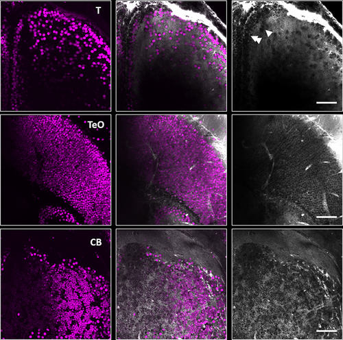
Visualization of cells with third harmonic generation (THG) microscopy in Casper line of adult zebrafish, Danio rerio. Nuclear-localized GCaMP-labeled neurons were simultaneously visualized via three-photon excitation (3PE) and THG in the telencephalon (T), optic tectum (TeO), and granule cell layer of the cerebellum (CB) (left column: 3PE, middle column: overlay of 3PE and THG, right column: THG). Closely packed cells along the outer ventricular margin of the telencephalon (top right) are pointed to by double white arrowheads and islands of closely packed cells located in area dorsalis are pointed out by single white arrowheads. All images collected with a pixel size of 0.26 μm. |