
Fig. 5
- ID
- ZDB-FIG-240523-67
- Publication
- Akbari et al., 2024 - Label-free, whole-brain in vivo mapping in an adult vertebrate with third harmonic generation microscopy
- Other Figures
- All Figure Page
- Back to All Figure Page
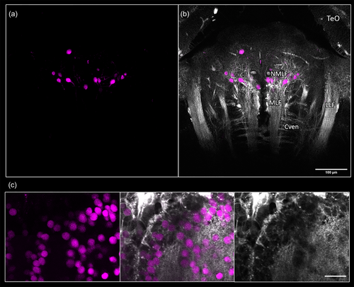
Demonstration of third harmonic generation (THG) microscopy as a navigation guide for in vivo fluorescence imaging. Anterior is to the top in all images. (a) Horizontal view of retrogradely labeled cells in the hindbrain of adult Danionella dracula following bulk injection of fluorescein into the spinal cord. (b) Overlay of THG microscopy with fluorescence image in (a). The lateral longitudinal fasciculus (LLF), medial longitudinal fasciculus (MLF), and axonal bundles of the ventral commissure (Cven) are clearly distinguishable in this image, providing landmarks for identification of the nucleus of the MLF (NMLF). TeO: optic tectum. (c) Nuclear-localized GCaMP-labeled neurons in Casper line of adult zebrafish were simultaneously visualized via three-photon excitation (3PE) and THG in the telencephalon of adult zebrafish, Danio rerio (left column: 3PE, middle column: overlay of 3PE and THG, right column: THG). Depending on the focal depth, the THG image corresponding to the fluorescently labeled neuron might appear faint for some cells. All images collected with a pixel size of 0.26 μm. |