
Fig. 4
- ID
- ZDB-FIG-240523-66
- Publication
- Akbari et al., 2024 - Label-free, whole-brain in vivo mapping in an adult vertebrate with third harmonic generation microscopy
- Other Figures
- All Figure Page
- Back to All Figure Page
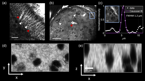
Visualizing individual cells with third harmonic generation (THG) microscopy in the adult brain of Danionella dracula. (a) Representative high magnification image from midbrain roof (optic tectum) showing closely packed cells (white arrowheads) and blood vessels (red arrowheads) within the periventricular gray zone. Scale bar indicates 20 μm. (b) Representative high magnification image from the pallium (area dorsalis of the telencephalon) showing layer of closely packed neurons along the outer ventricular margin (double white arrowheads) as well as individual neurons and clusters of closely packed neurons located more deeply (single white arrowheads); blood vessel is also indicated (red arrowhead). The anterior is toward top of the image. Scale bar indicates 50 μm. (c) Line profile of the area outlined in blue box in (b). A 5-pixel wide line is drawn on a small structure. The full width at half maximum (FWHM) of the profile is 1.2 μm, indicating that the technique can distinguish micrometer range features. Lateral (d) and axial (e) views of cell shadows in telencephalon obtained by THG microscopy. Images are minimum projection of column containing several cells acquired at 1 μm steps in z direction. Images collected with a pixel size of 0.26 μm (a, b, c) and 0.07 μm (d, e). |