|
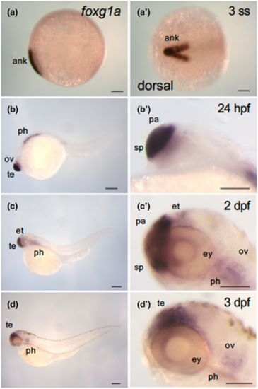
Dynamic expression of foxg1a during the course of zebrafish development. Expression of foxg1a was examined by WISH in wild-type embryos from the early somite stage through 3 dpf. (a–d, b′–d′) Lateral views of the entire bodies (a–d) and heads (b′–d′) with anterior to the left, dorsal to the top. (a′) Dorsal view, with anterior to the left. foxg1a expression was detected in the anterior neural keel from the two-somite stage (2-ss; Figure 1a, a′). At 24 hpf, foxg1a expression was detected in the telencephalon, including the preoptic area, optic vesicle, and pharyngeal arch. At 2 dpf, foxg1a expression was detected in the telencephalon, eyes, epithalamus, and pharyngeal arches. At 3 dpf, foxg1a expression was also found in the telencephalon, eyes, and pharyngeal arches. ank, anterior neural keel; ov, optic vesicle; pa, pallium; ph, pharyngeal arch; sp, subpallium; te, telencephalon. Scale bar, 100 μm.
|