
Fig. 3
- ID
- ZDB-FIG-240521-26
- Publication
- Chakraborty et al., 2024 - Inhibition of NF-κB-Mediated Proinflammatory Transcription by Ru(II) Complexes of Anti-Angiogenic Ligands in Triple-Negative Breast Cancer
- Other Figures
- All Figure Page
- Back to All Figure Page
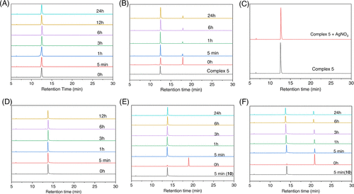
HPLC chromatograms of stability of complexes in 2:8 v/v DMSO and phosphate buffer (10 mM phosphate, 4 mM NaCl, pH 7.4) at different time intervals except (C) where the study was conducted in the presence of 1 equiv AgNO3 in 2:8 v/v DMSO/H2O. (A) Complex 5 showing the presence of a single peak, which corresponds to the monoaquated species as confirmed from the study in (C) where complex 5 is intentionally hydrolyzed instantaneously using 1 equiv of AgNO3 showing the same single peak at 12.5 min. (B) Complex 6 showing the monoaquated complex (12.5 min) and the intact 6 at 17.9 min. (D) Aquation of 10–12 showing the monoaquated complexes to be the sole species for 10 and 11, whereas 12 shows intact complex and monoaquated species in the beginning where the intact complexes decreases with time. Data plots from (E) and (F) show that both 11 and 12 convert into 10 in solution due to monoaquation. The time points indicated on the plot represent the injection times of these complexes. Mobile phase: mixture of water and acetonitrile (MeCN). Elution gradient: starting at 5% MeCN and gradually increasing to 95% MeCN over a 30 min duration. Flow rate: 1 mL/min. The reference point at 0 h corresponds to the stability of complexes in a 200 μM dimethyl sulfoxide (DMSO) solution. Detector UV channel = 254 nm. |