
Fig. 7
- ID
- ZDB-FIG-240517-33
- Publication
- Zhu et al., 2024 - Allelic heterogeneity of TTNtv cardiomyopathy can be modeled in adult zebrafish
- Other Figures
- All Figure Page
- Back to All Figure Page
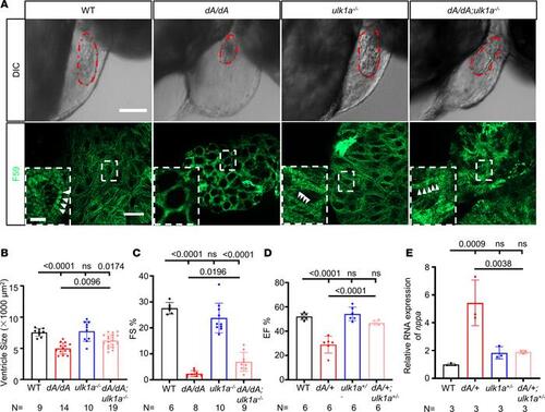
ulk1a mutants repair cardiac dysfunction in both adult and embryonic stages of ttntv-A mutants. (A) Immunostaining and differential interference contrast (DIC) images of 2 dpf embryos. Sarcomere structures were obtained by immunostaining with an anti-F59 (green) antibody in WT, dA/dA, ulk1a–/–, and dA/dA ulk1a–/–. Higher magnifications of the boxed areas are shown in the insets. White arrowheads indicate striated sarcomere structures. The embryonic ventricle is outlined by dashed red lines in the DIC images. The scale bar of the upper panel is 100 μm, the lower panel is 10 μm, and the inset is 2.5 μm. (B) Quantification of the ventricle area based on the area outlined by the dashed red lines in the DIC panels of Figure 7A. (C) Quantification of fractional shortening in embryonic hearts. (D) High-frequency echocardiography was performed on 6-month-old zebrafish to quantify EF%. (E) Evaluation of nppa gene transcript expression by quantitative real-time PCR in hearts from 3-month-old adult zebrafish. One-way ANOVA was used to compare multiple groups for each mutation. Data are presented as mean ± SD. |