FIGURE

Fig. 2

ID
ZDB-FIG-240515-35
Publication
Hao et al., 2024 - Somatic PDGFRB activating variants promote smooth muscle cell phenotype modulation in intracranial fusiform aneurysm
Other Figures
All Figure Page
Back to All Figure Page
Fig. 2

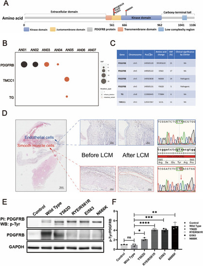
Somatic mutational landscape of FIAs. Structure of the protein encoded by PDGFRβ and the lollipop plots illustrate the location of the identified mutations in our cohort. The color of the boxes denotes different regions of the encoded protein. The coordinates represent the n-th amino acids (A). Somatic mutational landscape of 7 FIA samples with paired blood. The x-axis and y-axis specify the gene name and sample ID, respectively. The size of the circle represents the variant allele frequency. And the color denotes different types of mutations (B). Clinical significance of the detected somatic mutations according to the ClinVar database (C). The smooth muscle layer and endothelial layer in the FIA sections are dissected by laser-capture microdissection (LCM). PDGFRB.Y562D mutation is found in smooth muscle layer by Sanger sequencing (D). HBVSMCs with PDGFRB mutations identified by WES are constructed and these mutations are found to have a characteristic with gain-of-function by immunoprecipitation (E). The scatter bar graph is shown the relative density of immunoblot bands (normalized to those in cells transfected vector) about pTyr/PDGFRβ in cells expressing different PDGFRB mutations is shown in (F) (n = 3). ‘n’ represents three repeated experiments. The Student's t-test was employed to assess the differences in pTyr/PDGFRβ levels between different mutant groups and the Wild Type group, with Bonferroni correction for multiple comparisons. n values represent the number of repeated experiments. ns, no significant, **padj < 0.01, ***padj < 0.001

Expression Data

Expression Detail
Antibody Labeling
Phenotype Data

Phenotype Detail
Acknowledgments
This image is the copyrighted work of the attributed author or publisher, and ZFIN has permission only to display this image to its users. Additional permissions should be obtained from the applicable author or publisher of the image. Full text @ J. Biomed. Sci.