
Fig. 5
- ID
- ZDB-FIG-240515-11
- Publication
- Miladinovic et al., 2024 - A multistep computational approach reveals a neuro-mesenchymal cell population in the embryonic hematopoietic stem cell niche
- Other Figures
- All Figure Page
- Back to All Figure Page
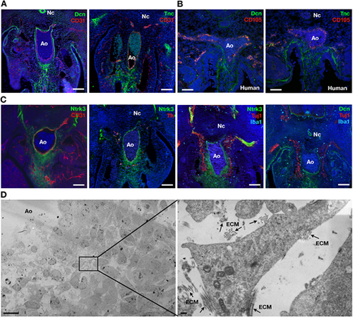
Morphological studies confirm the existence of a subaortic neuro-mesenchymal population. (A) Immunodetection of Dcn, Tnc and CD31 on E11.5 mouse embryo sections. (B) Immunodetection of Dcn, Tnc and CD105 on day 30 human embryo sections. (C) Immunodetection of Ntrk3, CD31, Th, Tuj1, Iba1 and Dcn on E11.5 mouse embryo sections. Unmerged images are shown in Fig. S7. (D) Transmission electron microscopy of E11.5 subaortic tissue. The image on the right is a magnification of the boxed area in the left panel. Arrows indicate areas of extracellular matrix (ECM). Ao, dorsal aorta; Nc, notochord. Scale bars: 100 µm (A,C); 50 µm (B); 10 µm (D, left); 200 nm (D, right) |