
Fig. 5
- ID
- ZDB-FIG-240513-27
- Publication
- Tanimoto et al., 2024 - Transgenic tools targeting the basal ganglia reveal both evolutionary conservation and specialization of neural circuits in zebrafish
- Other Figures
- All Figure Page
- Back to All Figure Page
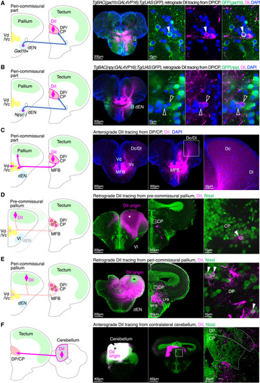
The npy-negative inhibitory dEN neurons project to the DP/CP thalamus and form the pallido-thalamo-pallial pathway (A) DiI tracing from the DP/CP in TgBAC(gad1b:GAL4VP16);Tg(UAS:GFP) fish retrogradely labeled gad1b+ dEN neurons. An inset in the leftmost panel shows the position of the right three panels. Filled arrowheads indicate retrogradely labeled gad1b+ dEN neurons. The leftmost panel is MaxIP. Scale bars, 200 μm (leftmost panel) and 10 μm (right three panels). (B) Same as (A) but in TgBAC(npy:GAL4VP16);Tg(UAS:GFP) fish. Open arrowheads indicate retrogradely labeled npy-negative dEN neurons. (C) DiI tracing from the DP/CP anterogradely labeled projection fibers in the MFB, the Dc/Dl pallium, and the Vc region. The same animal as in (B). Two peri-commissural sections are shown. An inset in the middle panel is magnified in the rightmost panel. MaxIP is shown. Scale bars, 200 μm (left two panels) and 50 μm (rightmost panel). (D) DiI tracing from the pre-commissural pallium retrogradely labeled thalamic neurons. An asterisk indicates DiI origin. An inset in the middle panel shows the position of the rightmost panel. An arrowhead indicates retrogradely labeled CP neurons. The left two panels are MaxIP. Scale bars, 200 μm (left two panels) and 10 μm (rightmost panel). (E) Same as (D), but from the peri-commissural pallium. Arrowheads indicate retrogradely labeled DP neurons. (F) DiI tracing from the cerebellum anterogradely labeled projection fibers in the contralateral DP/CP. An asterisk indicates DiI origin. An inset in the middle panel shows the position of the rightmost panel. The left two panels are MaxIP. Scale bars, 400 μm (left two panels) and 50 μm (rightmost panel). |