
Fig. 3
- ID
- ZDB-FIG-240513-25
- Publication
- Tanimoto et al., 2024 - Transgenic tools targeting the basal ganglia reveal both evolutionary conservation and specialization of neural circuits in zebrafish
- Other Figures
- All Figure Page
- Back to All Figure Page
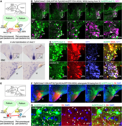
The indirect-pathway-targeted region Vl contains neurons expressing GP/GPe marker Nkx2.1 and projects to the direct-pathway-targeted region dEN (A) Illustration of coronal sections depicting WGA expression in the nkx2.1+ Vl neurons and trans-synaptic WGA transfer to the npy+ dEN neurons. (B) Different antero-posterior distributions of the nkx2.1+ neurons (AcGFP, green) and NPY+ neurons (NPY, magenta) in the five successive coronal sections (a–e). Insets in (a)–(e) show the positions of (a’)–(e’), focusing on the Vl-dEN region. Scale bars, 100 μm (a–e) and 20 μm (a’–e’). (C) ISH analysis of nkx2.1 in the Vl. Two successive coronal sections at the Vl are shown. The same animal as in Figure S1H. Scale bars, 200 μm (top panels) and 50 μm (bottom panels). (D) WGA tracing from the nkx2.1+ neurons and immunohistochemistry of AcGFP (green), WGA (red), and NPY (light blue). Upper and lower panels show the Vl (section a) and the dEN (section d), respectively. Arrowheads indicate NPY+ dEN neurons with WGA signals. Scale bars, 20 μm. (E) Illustration of coronal sections depicting anterograde DiI tracing from the Vl in TgBAC(nkx2.1:GAL4VP16);Tg(UAS:AcGFP-P2A-WGA) fish. (F) Five successive sections (f–j) of the DiI tracing from the Vl to the dEN, and multicolor imaging of AcGFP (green), DiI (red), and DAPI (blue). An inset in (e) shows the position of (G). MaxIP is shown. Scale bar, 100 μm. (G) Magnified views of the dEN (section j). Arrowheads indicate DiI and AcGFP double-positive fibers in the dEN, indicating direct projection from the nkx2.1+ Vl neurons to the dEN. Scale bar, 10 μm. |