|
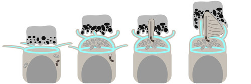
Diagram depicting stages of photoreceptor CP, IS and OS development in embryonic zebrafish. From left to right: CPs, the IS and the OS of zebrafish photoreceptors undergo distinct alterations during development. First on the left: no distinct IS is observed; photoreceptors feature tangential processes apically, an actin ring at the OLM and a flat RPE–IS interface. Next, the IS becomes prominent, outlined by an actin dome, and vertical processes (presumably CP precursors) appear, while the RPE–IS interface becomes rougher. Tangential processes originating near the OLM area are retained. Further along, a cilium, the future OS, emerges and enters the RPE pocket, with no processes adjacent to it. Finally, the cilium starts generating discs, the CPs associate with the new OS, and the IS becomes more rectangular in shape. Please note that the diagram does not accurately depict relative sizes of photoreceptors and RPE in order to highlight the apical region of the former.
|