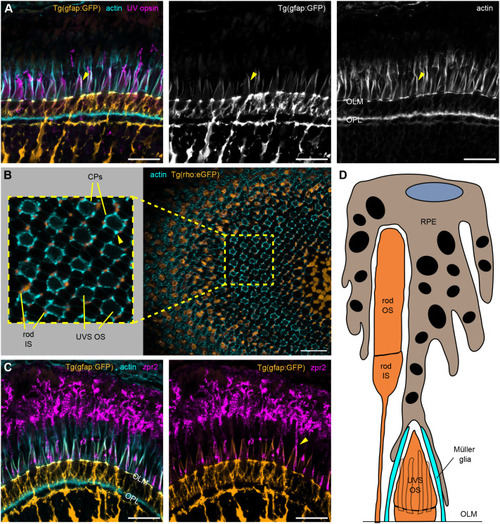
Zebrafish Müller glial and RPE protrusions enclose UVS cone OSs. (A–C) Confocal images of 1 mpf zebrafish retinal sections incubated with phalloidin (cyan) and UV opsin or zpr2 antibody (magenta). (A) Tg(gfap:GFP) zebrafish with Müller glia cell bodies highlighted by GFP (orange) show long glial processes above the OLM stretching alongside UVS cone OSs and colocalizing with thick actin bundles (arrowheads). (B) Sagittal section through Tg(rho:eGFP) retina with an enlarged area demonstrating rod ISs (orange) adjacent to thick actin bundles (arrowhead) surrounding UVS cones OSs. (C) RPE apical processes, stained with zpr2 antibody, extend towards the OLM and localize in close proximity to the apical Müller glial processes (arrowhead), as observed in Tg(gfap:GFP) retina. (D) A model illustrating the organization of supporting cells in the photoreceptor layer. UVS cones feature both Müller glial and RPE protrusions around the OS. Number of fish analysed: n=3 (A,C), n=5 (B). Scale bars: 20 µm.
|
