|
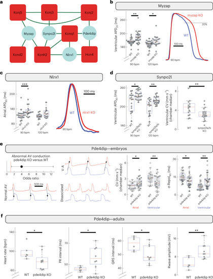
Functional evaluation of interactors shared across multiple channel networks. Four proteins that each interact with three different ion channels were functionally investigated. a, Interactions identified for Myzap, Nlrx1, Pde4dip and Synpo2l. Bait proteins are shown in red, interactors in light blue and interactions in green. b, Acute KO in zebrafish of the gene encoding myzap (n = 25 fish) resulted in prolonged ventricular APD (APD20; **P = 2.85 × 10⁻³; *P = 0.018 by two-sided Mann–Whitney U test; exemplar amplitude-normalized optical action potentials shown) compared to control siblings (WT, n = 25). c, KO of nlrx1 (n = 21) mainly affected atrial APD, with a greater effect size in late repolarization (APD80) and at slower heart rates (nWT = 21, ***P = 7.64 × 10⁻⁴ by idem; exemplar amplitude normalized optical action potentials shown). d, KO of synpo2la/b (n = 23) led to prolonged ventricular APD at multiple paced heart rates with greatest effect size late in repolarization (nWT = 17, *P = 0.014; **P = 5.04 × 10⁻³, idem) and reduced ventricular CV (sinus rhythm; **P = 0.015, idem). e, KO of pde4dip (n = 30) resulted in hearts more prone to abnormal AV conduction (nWT = 30, odds ratio and 95% CI shown; P = 0.029 by Fisher’s exact test; exemplars show normal atrioventricular conduction versus retrograde conduction or AV dissociation), decreased ventricular CV and increased spatial dispersion of ventricular repolarization (standard deviation of repolarization time across the chamber), but with inverted effects in the atrial chamber (intrinsic rhythm; CV: *P = 6.24 × 10⁻³; **P = 5.86 × 10⁻³; σ-Repol80: *P = 0.013; **P = 1.66 × 10⁻⁴, idem). This reduces the differential between the chambers, which was observed in pde4dip-deficient fish with both abnormal AV conduction (filled markers) and normal (open markers). This suggests episodic abnormal AV conduction resulting in electrical remodeling with persisting effects during periods of normal AV conduction. f, In adult zebrafish, pde4dip deficiency (n = 9) resulted in slower heart rate (*P = 0.049), longer PR and shorter QRS intervals (*P = 0.029 and P = 0.036, respectively), and greater R wave magnitude (**P = 0.0095, all by idem, nWT = 6). Each point in the box plots corresponds to an individual zebrafish. Box plots indicate 25th/50th/75th percentiles, while whiskers extend to the most extreme data within 1.5× of interquartile range beyond box limits. Source data
|