FIGURE

Fig. 1

ID
ZDB-FIG-240408-70
Publication
Dieris et al., 2024 - Calcium imaging of adult olfactory epithelium reveals amines as important odor class in fish
Other Figures
All Figure Page
Back to All Figure Page
Fig. 1

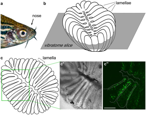
Slice preparation of adult zebrafish olfactory epithelium for calcium imaging. The position of the nose is shown by the white dotted oval (a). Schematic representation of the olfactory epithelium (b), lumen is up, individual lamellae and the median raphe are visible. The horizontal orientation and approximate height of the vibratome section plane is shown. En face view of the vibratome section (c), cross-sectioned lamellae and the median raphe are visible. In the schematic representation (c), the green rectangle corresponds approximately to the region shown in (c', c''). A representative vibratome section used for calcium imaging (bright field (c') and fluorescent signals (c'') after loading with Fluo4-AM, respectively)

Expression Data

Expression Detail
Antibody Labeling
Phenotype Data

Phenotype Detail
Acknowledgments
This image is the copyrighted work of the attributed author or publisher, and ZFIN has permission only to display this image to its users. Additional permissions should be obtained from the applicable author or publisher of the image. Full text @ Cell Tissue Res.