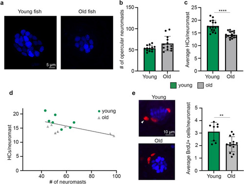
Young zebrafish have more hair cells and more dividing cells per lateral line neuromast. (a) Representative confocal images of opercular neuromasts from a young (left) and old (right) zebrafish. Hair cell nuclei were live-labeled with DAPI. Scale bar applies to both images. (b, c) Comparisons of superficial neuromast number (b) and hair cell number (c) on zebrafish opercula between young (green bars) and old (gray bars) fish. (b) There is no age difference in the number of superficial opercular neuromasts (Mann Whitney U test, p = 0.088). N = 13 fish/age class. (c) Young fish have significantly more hair cells per neuromast (2-tailed t-test, p < 0.0001). N = 16–17 fish/age class. (d) There is a negative relationship between neuromast number and hair cells/neuromast (R2 = 0.3716; n = 7 fish/age class). (e) Young fish have significantly more BrdU + cells per opercular neuromast (2-tailed t-test, p = 0.0069). N = 8–13 fish/age class. Confocal images on the left in panel (e) show DAPI + hair cell nuclei in blue and BrdU + cells in red. White arrowhead points to an example BrdU + cell and the scale bar applies to both images. Data represent the average of 10 neuromasts per fish for each dataset. **p < 0.01, ****p < 0.0001. Data are presented as mean ± 1 s.d. and dots represent individual fish.
|
