|
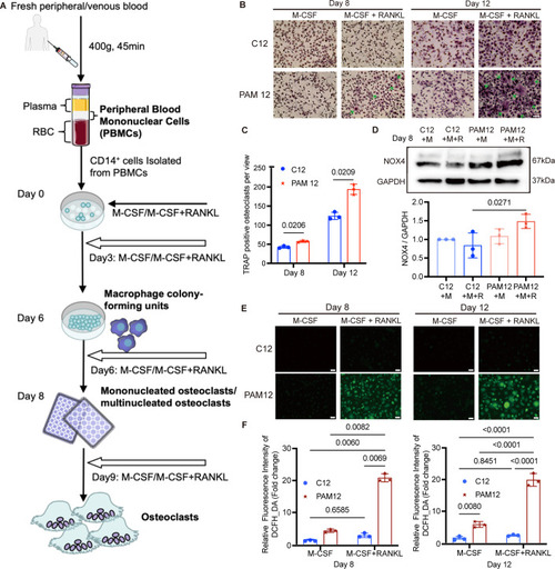
PAM12-derived osteoclasts show enhanced differentiation and increased ROS generation activity compared to osteoclast-derived cells from healthy control. (A) Schematic illustration of isolation of osteoclasts from peripheral blood. More details are shown under “Methods”. (B) Osteoclasts were differentiated with colony-stimulating factor (M-CSF/M) and receptor activator of nuclear factor κB ligand (RANKL/R). Tartrate-resistant acid phosphatase (TRAP) staining (violet-labeled) was used to mark differentiated osteoclast (> 3 nuclei). PAM12-derived cells show a higher number of differentiated osteoclasts (marked by arrows) compared to cells derived from a healthy control (C12). (C) The number of TRAP-positive osteoclasts per view was counted blindly by 2 persons. N = 3. (D) Western blot detected the NOX4 protein expression in differentiated osteoclasts derived from PAM12 and C12 on Day 8, and GAPDH was used as a loading control. N = 3. (E) ROS probed by DCFH-DA in cells from PAM12 was significantly higher compared to control cells at both 8 and 12 days after differentiation with M-CSF and RANKL. Representative images are shown. N = 3. (F) The average fluorescence intensity was quantified by ImageJ software at two time points. N = 3. Data information: scale bars = 100 μm. N = biological replicates. (C, D, F) Error bars in figures represent mean ± SEM. One-way ANOVA with Tukey’s multiple comparisons test was used to determine significant differences. Source data are available online for this figure.
|