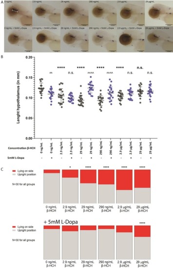
Whole-mount in situ hybridization analysis with TH1 probe in a 3 days post fertilization embryo, exposure to β-HCH, additional L-Dopa treatment, and distribution of larval posture. A) Whole-mount in situ hybridization analysis with TH1 probe in a 3 days post fertilization embryo. The hypothalamus is indicated in the upper left panel by a white bar. Upper panels are representative larvae treated with β-HCH at the indicated concentrations. Lower panels are representative images of larvae treated 5 mM L-Dopa in addition to indicated concentrations of β-HCH. Scale bars are 0.25 mm. B) Length of the hypothalamus of 3 days post fertilization embryos exposed to indicated concentrations of β-HCH, with (blue) or without (grey) additional L-Dopa treatment. Error bars represent S.E.M., significance was calculated using one-way ANOVA with Dunnett’s post-test. The ‘*’ symbol is used to indicate significance of difference compared to the 0 ng/mL group. The ‘^’ is used to indicate significance of differences between groups treated with the same concentration of β-HCH, with or without 5 mM L-Dopa. C) Distribution of larval posture at 3 days post fertilization after exposure to β-HCH in early developmental stages, without (upper panel) or with (lower panel) simultaneous exposure to 5 mM L-Dopa. Significance was calculated using Chi-square test. (For interpretation of the references to colour in this figure legend, the reader is referred to the web version of this article.)
|
