
Fig. 8
- ID
- ZDB-FIG-231220-93
- Publication
- Palsamy et al., 2023 - Microglial depletion after brain injury prolongs inflammation and impairs brain repair, adult neurogenesis and pro-regenerative signaling
- Other Figures
- All Figure Page
- Back to All Figure Page
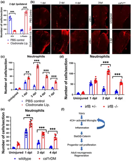
Persistence of neutrophils when microglia are ablated in the setting of brain injury. (a) Quantification of microglia (4C4) and leukocytes (L-Plastin) in the ipsilesional hemi-telencephala of control or clodronate liposome injected fish at 2 dpl. (b) Confocal images of fish telencephala from PBS control and clodronate treated groups at 1–4 dpl, and irf8+/− (control), irf8−/− and csf1rDM groups at 2 dpl, all immunolabeled for an Mpx-mcherry reporter (neutrophils). (c–e) Quantification of Mpx reporter-positive neutrophils from control, clodronate treated, irf8 and csf1rDM mutant brains. (f) Proposed model of the microglia-induced signaling pathway during injury-induced telencephalic regeneration in zebrafish. ***p < .0001; **p < .001; *p < .01 by t-test for panels (a, c, d, and e). Scale bars = 100 μm, and the error bars indicate SD. |