
Fig. 5
- ID
- ZDB-FIG-231220-90
- Publication
- Palsamy et al., 2023 - Microglial depletion after brain injury prolongs inflammation and impairs brain repair, adult neurogenesis and pro-regenerative signaling
- Other Figures
- All Figure Page
- Back to All Figure Page
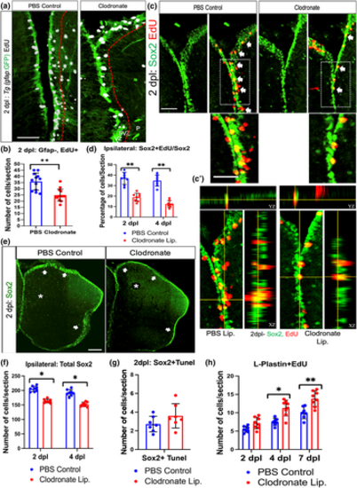
Microglial ablation impairs injury-induced amplifying intermediate progenitor cell (aIPC) expansion. (a) Confocal images of midline telencephalic sections from clodronate and control liposome injected fish double labeled for a gfap-driven GFP reporter (green) and EdU (white) at 2 dpl. The dashed red lines divide the VZ + SVZ (left of the lines) from the parenchyma (P). (b) Quantification of gfap-driven GFP reporter-negative/EdU-positive cells at 2 dpl in the VZ plus SVZ. **p < .001 by t-test. (c) Confocal images of double labeling for Sox2 (green) and EdU (red) at 2 dpl in the VZ + SVZ at the midline telencephalon. Arrows indicate double-positive cells, and the boxed areas are shown at higher magnification below. (c′) Orthogonal views of single optical sections from z-stack images of Sox2 and EdU labeling. (d) Quantification of percentages of Sox2+ cells that co-labeled with EdU in the ipsilesional telencephalon at 2 and 4 dpl. **p < .001 by t-test. (e) Sox2 immunolabeling of lesioned control liposome and clodronate liposome treated fish telencephala at 2 dpl. Arrows indicate labeled progenitor cells in the VZ/SVZ and asterisks denote the damaged regions. (f) Quantification of Sox2-labeled cells in control and clodronate injured brains at 2 and 4 dpl. *p < .01 by t-test. (g) Quantification of Sox2/TUNEL co-labeled cells at 2 dpl in control and clodronate groups. (h) Quantification of L-Plastin and EdU double-labeled cells at 2, 4, and 7 dpl in control and clodronate injected groups revealed significantly increased double-labeled cell numbers in the clodronate group at 4 and 7 dpl. **p < .001; *p < .01. Scale bar = 100 μm for all image panels except 50 μm for the higher magnification images in panel (c); error bars indicate SEM. |