
Fig. 6
- ID
- ZDB-FIG-231220-91
- Publication
- Palsamy et al., 2023 - Microglial depletion after brain injury prolongs inflammation and impairs brain repair, adult neurogenesis and pro-regenerative signaling
- Other Figures
- All Figure Page
- Back to All Figure Page
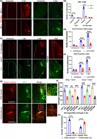
Reduced injury-induced proliferation after genetic depletion of microglia, and persistent macrophages after clodronate suppression of microglia. (a) Immunostaining for 4C4 (microglia, red) and EdU (proliferating cells, green) in irf8+/− versus irf8−/− fish telencephala with or without injury at 2 dpl. (b) Quantification of microglia and EdU-positive cells in irf8 mutants and controls without injury or at 2 dpl. (c) Staining for 4C4 (red) and EdU (green) in csf1rDM mutant (left panels) and wildtype (right panels) brains with or without injury. Dashed lines indicate the midline in (a and c). (d) Quantification of microglia in csf1rDM and wildtype brains. (e) EdU-positive cell quantification in csf1rDM and wildtype brains. (f) Quantification of cells double-labeled for Gfap/EdU or Sox2/EdU in mutant fish (irf8−/− and csf1rDM lines) and fish treated with PLX3397 compared to control lines (irf8+/−, DMSO and wildtype) at 2 dpl. (g) Double immunolabeling of telencephala for mCherry to label mpeg1-positive cells (red) from mpeg1:mcherry reporter fish and 4C4 (green) for microglia in control and clodronate injected fish. The images are ipsilesional, and the dotted boxes are shown at higher magnification on the right. (h) Quantification of microglia (mpeg+, 4C4+) and macrophages (mpeg+, 4C4−) in control and clodronate brains at 2 dpl. ***p < .0001 for all graphs. Scale bars = 100 μm for all image panels, and the error bars indicate SEM. |