|
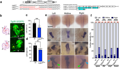
cygb2 mutant phenotype presents organ laterality defects.\ a CRISPR/Cas9 mediated genome editing of cygb2. Two different gRNA were targeted to exon 1 (denoted by red text) and resulted in 4 bp and 1 bp frame shift mutations (beginning in the blue shaded region of the predicted protein structure) named cygb2801a and cygb2801b, respectively. The eight globin protein helices (labeled A-H) are represented by boxes, with out-of-frame amino acids shaded blue. b Whole mount 3D confocal projections (right) of wt and cygb2pt801atg(fli1eGFPy1) hearts at 4 days post fertilization (dpf) with schematic (left) representing the heart morphology and direction of blood flow. V – ventricle, A – atrium, Y – yolk. Scale bar = 20 µm. c, d Quantification of the percentage of embryos with a left-sided heart loop in cygb2801a and cygb2801b. Means are ± SD (n = 6–7, each n representing an independent experiment consisting of 50 embryos). Student’s t test, two-tailed, **P < 0.01, ***P < 0.001. e Representative in situ hybridization images of the cygb2801a laterality phenotype. southpaw (spaw), 16 somites, dorsal view; lefty2 (lft2), 22 h post fertilization (hpf), dorsal view; myosin light chain 7 (myl7), 96 hpf, ventral view; and foxa3, 2 dpf, dorsal view. Green arrow heads indicate the liver and blue arrow heads point to the pancreas. Scale bars = 100 μm. f Quantification of the percentage of embryos with right, straight/bilateral or left sided expression of spaw, lft2, mly7 or foxa3. The total number of embryos analyzed is shown in red above the graph. The Chi-squared test was used to determine statistical significance. Source data are provided as source data file.
|