FIGURE

Figure 1.

ID
ZDB-FIG-231212-42
Publication
Singh et al., 2023 - NMJ analyser: a novel method to quantify neuromuscular junction morphology in zebrafish
Other Figures
All Figure Page
Back to All Figure Page
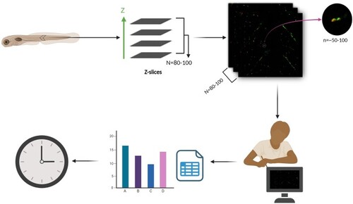
Figure 1.

Manual analysis of NMJ morphology is time-consuming and prone to bias. Confocal imaging of zebrafish NMJs, stained with presynaptic marker SV2a and postsynaptic marker α-bungarotoxin (α-BTX), entails acquisition of 80–100 z-slices (N). Each z-slice contains an estimated 50–100 synaptic puncta (n). The manual quantification of individual puncta within separate channels (2) and the assessment of their co-localization necessitate a substantial investment of time and effort, particularly when analyzing multiple samples representing diverse experimental conditions and varying time points. Moreover, limitations in the resolution of confocal microscopy introduce the possibility of under or overestimation of synaptic puncta counts.

Expression Data

Expression Detail
Antibody Labeling
Phenotype Data

Phenotype Detail
Acknowledgments
This image is the copyrighted work of the attributed author or publisher, and ZFIN has permission only to display this image to its users. Additional permissions should be obtained from the applicable author or publisher of the image. Full text @ Bioinformatics