
Fig. 5
- ID
- ZDB-FIG-231110-36
- Publication
- Brown et al., 2023 - Genetically Encoded Aminocoumarin Lysine for Optical Control of Protein-Nucleotide Interactions in Zebrafish Embryos
- Other Figures
- All Figure Page
- Back to All Figure Page
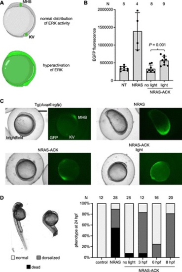
Photoactivation of a RASopathy mutant NRAS. (A) Expected distributions of EGFP in tg(dusp6:egfp) at bud stage (10 hpf). EGFP expression is indicative of ERK activity. At 10 hpf, expression is restricted to Kupffer’s vesicle (KV) and the mid-hindbrain boundary (MHB). (B) EGFP fluorescence was quantified from embryos at 10 hpf for each condition. Irradiation was performed at 6 hpf. Bars represent mean, and error bars represent standard deviation. An unpaired two-tailed Student’s t-test was performed between the two samples indicated. NT = nontreated embryos. (C) Representative images of Tg(dusp6:egfp) embryos for each condition. Scale bar = 0.5 mm. (D) Embryos were irradiated at the specified timepoint and scored for dorsalization defects at 24 hpf. Representative images are shown on the left. N = number of embryos. |