
Fig. 3
- ID
- ZDB-FIG-231110-34
- Publication
- Brown et al., 2023 - Genetically Encoded Aminocoumarin Lysine for Optical Control of Protein-Nucleotide Interactions in Zebrafish Embryos
- Other Figures
- All Figure Page
- Back to All Figure Page
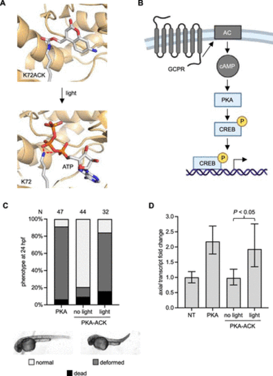
Photoactivation of Protein Kinase A (PKA). (A) Structural representation of the caged PKA nucleotide-binding pocket before and after light exposure. Red lines indicate hydrogen bonds (models generated from PDB: 4WB5). (B) The caPKA mutant is insulated from upstream-coupled protein receptor (GPCR) and adenylate cyclase (AC) interactions. (C) Embryos expressing PKA-ACK were irradiated at 4 hpf and imaged at 24 hpf. N = number of embryos. (D) Reverse transcription-quantitative polymerase chain reaction (RT-qPCR) measurement of the axial transcript. Bars represent mean, and error bars represent standard deviation from three independent pools of 50 embryos. An unpaired two-tailed Student’s t-test was performed between the two samples indicated. NT = nontreated embryos. |