
Fig. 2
- ID
- ZDB-FIG-231110-33
- Publication
- Brown et al., 2023 - Genetically Encoded Aminocoumarin Lysine for Optical Control of Protein-Nucleotide Interactions in Zebrafish Embryos
- Other Figures
- All Figure Page
- Back to All Figure Page
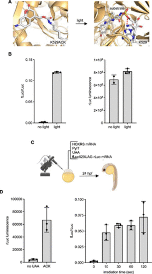
Incorporation of ACK into luciferase. (A) Structural representation of caging the fLuc active site with ACK (models generated from PDB: 4G36). Red lines indicate hydrogen bonding between K529 and luciferin–adenylate (here, a sulfate analogue as the substrate). (B) Incorporation of ACK into a dual-luciferase reporter and photoactivation of fLuc activity with 405 nm light in mammalian cells. Bars represent mean and error bars represent standard deviations of biological duplicates (no light) or triplicates (light). (C) Incorporation of ACK into a dual-luciferase reporter in zebrafish embryos is readily accomplished through injection of the mRNAs, tRNA, and UAA. (D) Luciferase incorporation and photoactivation assays. Embryos were irradiated with a 405 nm LED for the indicated amount of time. Bars represent mean, and error bars represent standard deviation from three pooled lysates of 4 embryos each. |