
Fig. 7
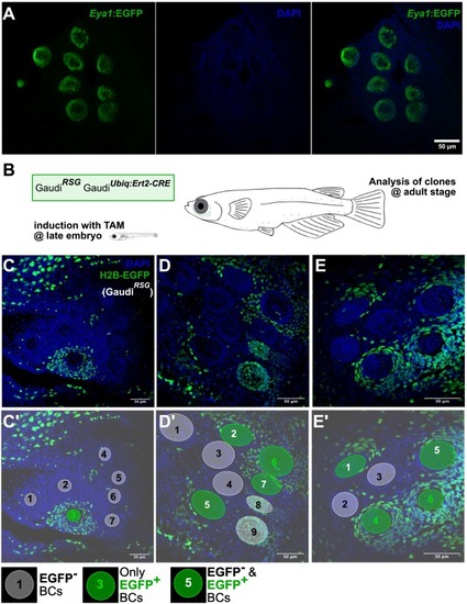
Niche expansion during post-embryonic life. (A) Confocal image of a Tg(Eya1:EGFP) adult medaka fish counterstained with DAPI, displaying 9 neuromasts arranged in the so-called caudal neuromast cluster. (B) Clone-induction strategy. Fish were treated with tamoxifen during late embryonic stage and imaged at adult stage. (C-E’) Examples of caudal neuromast clusters of recombined Gaudi fish. Clones over the border cell compartment are big and typically cover an entire neuromast (C), neighbor neuromasts (E) or a continuous, big portion of a neuromast (D). C’, D’, E’ are annotated versions of C, D, E and indicate EGFP+, EGFP− and hybrid niche compartments. Scale bar is 50 μm. |