
Fig. 6
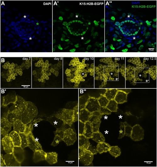
Sequential recruitment of niche cells during neuromast formation. (A) Confocal image of a Tg(K15:H2-EGFP) 10dfp larva counterstained with DAPI. White asterisks indicate EGFP− border cells, green asterisk indicates an EGFP+ border cell. (B) Time-lapse imaging of the same clone on a medaka embryo injected with the K15:mYFP construct. Gray circle in B indicates the position of the forming neuromast, white asterisks in B’, B” indicate border cells already converted where YFP signal decreases between 11 and 12.5 dpf, yellow asterisk in B’, B” indicates a newly recruited border cell during the same time frame. Scale bar is 10 μm in A-A”, B’, B” and 50 μm in B. |