
Fig. 5
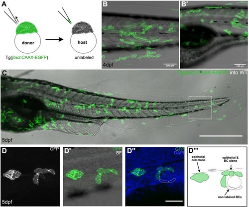
Multiclonal recruitment of border cells in zebrafish. (A) Schematic representation of blastula transplantation in zebrafish, taking a few cells from the donor Tg(βact:CAAX-EGFP) into an unlabeled wildtype host. (B-B´) Live images of 4 dpf chimeric zebrafish larvae, with EGFP+ clones along the body. (C) Fixed chimeric zebrafish larva at 5dpf showing EGFP+ clones that include epithelial/border cells. (D) Zoom-in of the chimeric zebrafish larva (squared in C). The clone including epithelial and border cells (D, D’) does not cover the entire niche compartment of the neuromast (D”) as depicted in the schematic representation (D”’). Scale bar is 100 μm in B, B’, 500 μm in C and 50 μm in D-D”. |